Cardiac Electrophysiological Imaging and Mapping
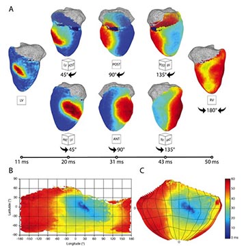
CVET develops high-resolution optical, electrical, and computational approaches to map cardiac activation, conduction, repolarization, and arrhythmia dynamics. These technologies include optical mapping, flexible electrode arrays, electro-optical interfaces, and advanced signal-processing methods that enable detailed characterization of normal and diseased cardiac function. The resulting information can guide the development of pacing, ablation, and other rhythm-management strategies. Vascular Imaging and Functional Assessment
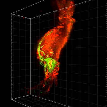
CVET applies and develops imaging methods to assess vascular anatomy, perfusion, remodeling, inflammation, and tissue recovery. Relevant approaches may include ultrasound, Doppler and laser-based perfusion imaging, optical and fluorescence imaging, molecular imaging, and three-dimensional vascular reconstruction. These tools support the study of ischemic disease, aneurysm progression, vascular implants, and regenerative therapies.